Ultrasensitive measurement of brain penetration mechanics and blood vessel rupture with microscale probes
Dr. Wu, Yu-Wei - May, 2026
Implanting microscale neural probes is central to neuroscience research and emerging brain-computer interfaces, yet the mechanical process by which tiny probes pass through living brain tissue has remained difficult to quantify. In this study, Dr. Yu-Wei Wu’s team at the Institute of Molecular Biology, Academia Sinica, and collaborators at Stanford University developed an ultrasensitive force-measurement system and combined it with real-time fluorescence and two-photon microscopy to directly observe probe insertion in the mouse brain. A portion of this research was conducted at the Center for Nano Science and Nanotechnology at National Sun Yat-sen University. By systematically comparing probes 7.5 to 100 micrometers in diameter, the team identified the pia as the main mechanical barrier during implantation. Puncture force and tissue compression increased with probe diameter, whereas the force required after crossing the pia remained nearly constant with depth. Live imaging further revealed a critical size regime below about 25 micrometers: in this range, blood vessels could be displaced rather than captured and torn, enabling insertion without visible bleeding. These findings provide quantitative design principles for safer, lower-trauma, high-density neural interfaces.
微型神經探針是腦科學研究與腦機介面技術的重要工具,但探針插入活體腦組織時需要多大力量,以及會如何影響血管與周邊組織,過去一直缺乏精確量化。本研究由中央研究院分子生物研究所吳玉威助研究員與碩士班研究生黃嵩文,和史丹佛大學團隊合作完成。研究團隊整合超高靈敏度力量量測平台,並結合即時螢光顯微與雙光子顯微成像,直接觀察不同尺寸探針插入小鼠腦部的過程;部分研究成果於國立中山大學奈米中心完成。系統性比較直徑 7.5 至 100 微米的探針後,研究團隊發現,腦表面的軟腦膜是植入時最主要的機械障礙;探針越細,穿刺所需力量越小,造成的組織壓縮也越少。探針穿過軟腦膜後,繼續深入腦組織所需力量幾乎不再隨深度增加。更重要的是,當探針直徑小於約 25 微米時,血管傾向被推移而非被勾住並撕裂,插入過程中未見可見出血。這項成果為設計更安全、低創傷、高密度的神經植入裝置提供了量化依據。
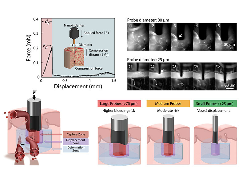
探針尺寸與血管損傷風險的概念模型。較大探針較容易勾住並拉伸鄰近血管,增加血管破裂與出血風險;隨探針直徑縮小,血管較可能被推移而非被撕裂。本研究中,直徑小於約 25 微米的探針插入時未觀察到可見出血。